Minimally invasive localization of breast lesions

The use of clips for the localization of breast lesions, instead of traditional wire hook placement, represents a significant advancement in the management of breast cancer. This shift in practice brings several clinical and practical benefits, improving both patient outcomes and surgical efficiency.

What is minimally invasive breast lesion localization?

Minimally invasive localization is a technique used to accurately mark a lesion detected via mammography or ultrasound.

Unlike traditional wire localization which involves inserting a wire into the breast on the day of surgery this method uses a metal clip placed at the time of biopsy. The clip remains in place until surgery, allowing for precise targeting of the lesion and more efficient removal.

How the localization procedure is performed at MIB Center Paris

During the biopsy, performed under ultrasound or mammographic guidance, a small metal clip is inserted at the exact location of the abnormality. This marker becomes visible during future imaging and acts as a guide during surgery.

On the day of surgery, a pre-operative localization scan is performed using a probe guided by ultrasound or mammography. This step allows the surgeon to accurately locate the lesion and adjust the incision size to better preserve surrounding healthy tissue.

The procedure typically lasts 20 to 30 minutes per lesion.

Benefits of pre-operative clip localization

More precise targeting and simplified procedure

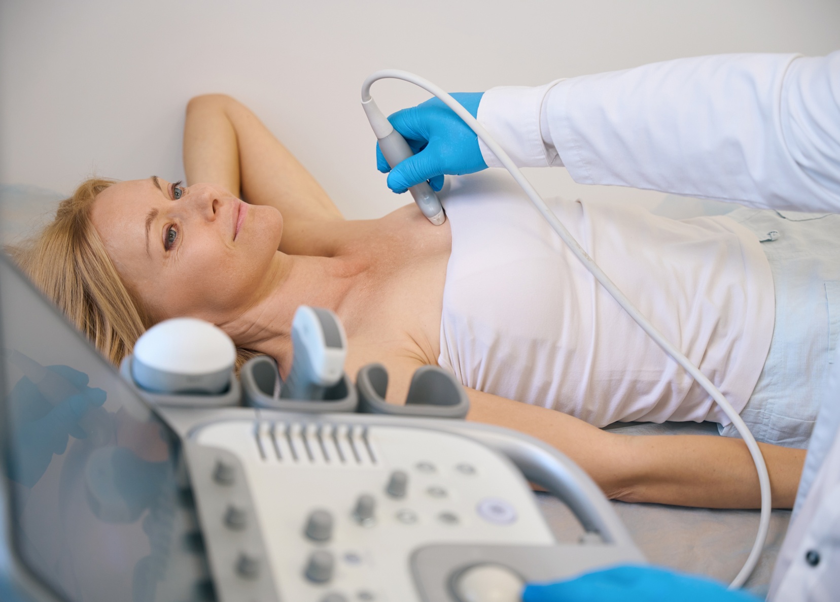
One of the main reasons to prefer clips over wires is the greater precision and simplified workflow they provide. The clip is placed with pinpoint accuracy during the biopsy, using imaging guidance.

This method allows for:

  • Highly accurate localization of the lesion before surgery
  • Easier, more effective tumor excision for the surgeon
  • Improved likelihood of complete tumor removal, reducing the risk of reoperation

Unlike the wire, which is inserted on the same day as surgery and can be uncomfortable or even painful, the clip is placed days or weeks before surgery, minimizing stress on the patient.

Improved comfort for patients

A key benefit of this method is patient comfort:

  • The wire, often a source of anxiety and discomfort, is replaced by a small, pain-free clip placed during biopsy
  • Patients arrive at the operating room calmer, without the added stress of same-day localization
  • Faster recovery with less post-operative pain

Better stability and higher accuracy

The clip remains stable and does not shift, ensuring precise localization during surgery. In contrast, wires may move or dislodge, complicating the procedure and compromising accuracy.

The fixed position of the clip supports a more effective, reliable intervention.

Better surgical planning and organization

Using clips also improves surgical scheduling and logistics. Traditional wire localization requires coordination between radiology and surgery on the same day, whereas clip placement allows for greater flexibility and smoother operating room workflows.

This results in:

  • More efficient use of hospital resources
  • Shorter waiting times for patients

Fewer risks and complications

Complications associated with wire localization such as bleeding, infection, or procedure-related pain are greatly reduced with clip use. This leads to faster, simpler recovery and improved patient experiences.

Clip localization: a step forward in breast cancer surgery

In conclusion, clip localization of breast lesions offers multiple advantages over traditional wire-guided methods. It enhances surgical precision, patient safety, and comfort, while also optimizing hospital resources.

This shift toward a less invasive, more reliable technique reflects the ongoing commitment of modern medicine to improving both the quality of care and patient outcomes in breast cancer treatment.

exchanges with web users

exchanges with web users